KPV peptide: complete guide to the anti-inflammatory α-MSH tripeptide
KPV is a three-amino-acid peptide, lysine-proline-valine (Lys-Pro-Val), matching positions 11 to 13 of alpha-melanocyte-stimulating hormone (α-MSH). This small fragment keeps much of the anti-inflammatory activity of the full hormone, without its pigmenting effect. Since the late 1980s it has been studied in models of fever, dermatitis, colitis and airway inflammation. Its best-known feature comes from a 2008 study in Gastroenterology: KPV enters intestinal cells through the peptide transporter PepT1, which explains why oral KPV is active in mice.
This guide gathers the published data: origin, mechanism, colitis studies, colon-targeted nanoparticles, skin data, oral versus injectable KPV, comparison with BPC-157 and Melanotan 2, reconstitution and regulatory status. One point first: no human clinical trial of KPV has been published. Every result cited here comes from cell cultures and animal models. The products mentioned are intended exclusively for in vitro and in vivo research use (RUO), not for human use.
Origin: the C-terminal fragment of α-MSH
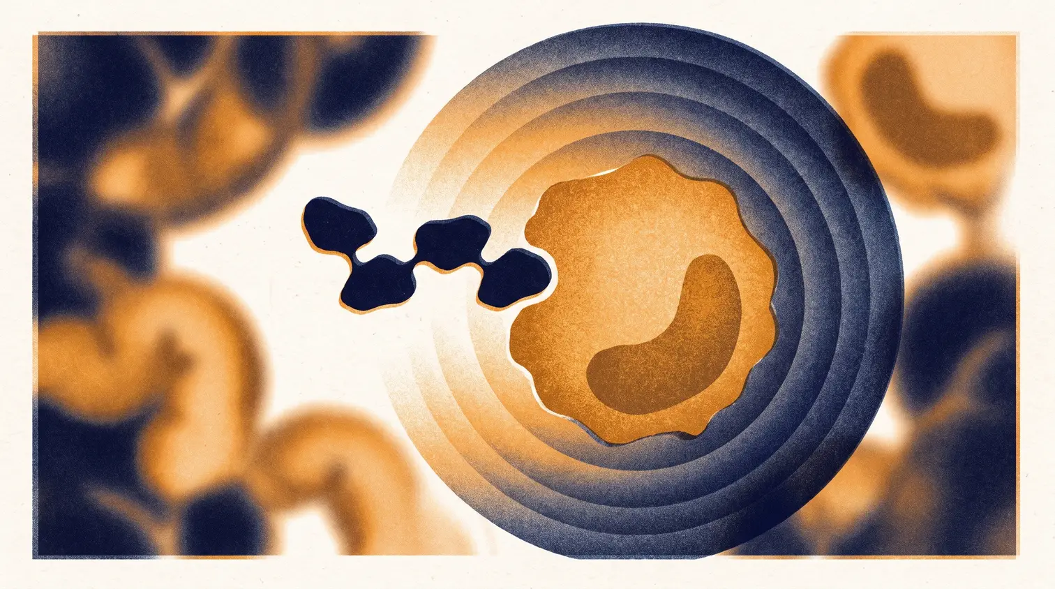
α-MSH is a 13-amino-acid hormone (Ser-Tyr-Ser-Met-Glu-His-Phe-Arg-Trp-Gly-Lys-Pro-Val) cleaved from pro-opiomelanocortin (POMC). It is best known for pigmentation, through the MC1R receptor on melanocytes. It is also present in barrier organs such as the skin and the gut, where it takes part in host defence and in the control of inflammation.
In the 1980s, J. M. Lipton's group in Dallas (Texas) showed that α-MSH lowers fever, and that this antipyretic activity appeared to sit in its C-terminal tripeptide, Lys-Pro-Val. In 1989, Hiltz and Lipton (FASEB Journal) tested this fragment, α-MSH(11-13), on picryl chloride-induced ear swelling in mice, a contact dermatitis model. The tripeptide reduced swelling in a dose-related fashion, compared with saline and a large dose of corticosteroid. It was one of the first published demonstrations of a general anti-inflammatory activity of KPV.
Reviews from the Münster group (Luger and Brzoska, Annals of the Rheumatic Diseases, 2007; Brzoska et al., Endocrine Reviews, 2008) sum up the interest: most of the anti-inflammatory activity of α-MSH can be attributed to its C-terminal tripeptide, which lacks the hormone's pigmentary action. That pigmentary effect was precisely what held back full-length α-MSH in inflammatory disease.
Molecular identity
| Parameter | Value |
|---|---|
| Common names | KPV, Lys-Pro-Val, α-MSH(11-13) |
| Sequence | H-Lys-Pro-Val-OH (L-lysyl-L-prolyl-L-valine) |
| Molecular formula | C16H30N4O4 |
| Molecular weight | 342.43 g/mol (monoisotopic 342.23 Da) |
| CAS number | 67727-97-3 (PubChem CID 125672) |
| Character | Highly hydrophilic (computed XLogP -3.5), positively charged below pH 7 |
In native α-MSH the terminal valine is amidated; PubChem lists the free-acid form, and some studies use Ac-Lys-Pro-Val-NH2 (Chavatte et al., 2001). A close derivative, KdPT (Lys-D-Pro-Thr), matches residues 193-195 of interleukin-1β and was developed as another anti-inflammatory tripeptide by the same research school.
Anti-inflammatory mechanism of action
Independent of melanocortin receptors
α-MSH acts on melanocortin receptors through its core His-Phe-Arg-Trp motif. KPV does not contain this motif. Getting, Schiöth and Perretti (Journal of Pharmacology and Experimental Therapeutics, 2003) compared it with α-MSH, the His-Phe-Arg-Trp core and Melanotan II in crystal-induced peritonitis in mice. Given systemically, KPV reduced neutrophil influx; the effect was not blocked by the MC3/MC4 antagonist SHU9119, KPV did not raise macrophage cyclic AMP, and the effect persisted in mice with a non-functional MC1R. The authors conclude that KPV is unlikely to act through melanocortin receptors and more likely works by inhibiting IL-1β functions.
The debate is not closed: Elliott et al. (Journal of Investigative Dermatology, 2004) observed KPV-induced calcium signals in cells transfected with MC1R. According to Brzoska et al. (2010), the exact signalling mechanism of KPV remains unknown.
NF-κB blockade
The best-documented target is the transcription factor NF-κB, which drives the production of many inflammatory cytokines. In Dalmasso et al. (2008), nanomolar KPV (most experiments used 10 nM) inhibited NF-κB and MAP kinase activation in human intestinal epithelial cells (Caco2-BBE, HT29-Cl.19A) and T cells (Jurkat), and reduced pro-inflammatory cytokine secretion.
Land (International Journal of Physiology, Pathophysiology and Pharmacology, 2012) refined the mechanism in human bronchial epithelial cells (16HBE14o-). KPV enters the nucleus, stabilises the inhibitor IκBα and blocks nuclear translocation of the NF-κB p65 (RelA) subunit. Competition assays point to an interaction with the importin-α3 binding site on p65. The result is a dose-dependent fall in NF-κB activity, MMP-9 activity and secretion of the chemokines IL-8 and eotaxin.
The PepT1 transporter
PepT1 (SLC15A1) is a di- and tripeptide transporter normally expressed in the small intestine. It is scarce in the healthy colon but induced in the colon during inflammatory bowel disease. Dalmasso, Charrier-Hisamuddin, Nguyen, Yan, Sitaraman and Merlin (Gastroenterology, 2008, Emory University) showed that KPV is a PepT1 substrate. Using tritiated KPV, they measured an apparent affinity constant (Km) of about 160 µM in Caco2-BBE cells and about 700 µM in Jurkat cells. At 100 µM, KPV reduced uptake of the reference substrate Gly-Sar by about 45%.
The consequence: the more inflamed the colon, the more PepT1 it expresses and the more KPV it takes up. Viennois et al. (Cellular and Molecular Gastroenterology and Hepatology, 2016) confirmed this genetically: in PepT1-knockout mice, KPV lost its protective effect.
Pharmacokinetics: what is known and what is not
No pharmacokinetic study of KPV has been published in humans: no plasma half-life, no measured oral or subcutaneous bioavailability. The available evidence is indirect.
- Intestinal absorption: PepT1-mediated uptake is shown in vitro and in mice (Dalmasso 2008, Viennois 2016).
- Skin penetration: Pawar et al. (Journal of Pharmaceutical Sciences, 2017) detected no passive permeation across human skin (detection limit 0.01 µg/mL). Microneedles raised the flux to 4.4 µg/cm²/h, and iontophoresis combined with microneedles increased it 35-fold over microneedles alone.
- Degradation: under stress (acid, alkali, hydrogen peroxide), KPV mainly degrades into Lys-Pro diketopiperazine (Pawar et al., Biomedical Chromatography, 2015).
Rapid clearance by peptidases is expected for an unmodified tripeptide, but it has not been quantified. Hence the delivery systems developed since.
Colitis studies in animals
Experimental colitis is where KPV has been studied most. The two main models are dextran sodium sulfate (DSS) colitis, which destroys the epithelial barrier, and TNBS colitis, which triggers an immune response.
| Study | Model | KPV administration | Main result |
|---|---|---|---|
| Dalmasso et al., Gastroenterology, 2008 | Mice, 3% DSS colitis (8 days) and TNBS 150 mg/kg | 100 µM in drinking water | Colonic MPO activity down about 50% (DSS) and 30% (TNBS); lower IL-1β, IL-6, TNF-α and IFN-γ (TNBS) |
| Kannengiesser et al., Inflammatory Bowel Diseases, 2008 | Mice, DSS colitis and CD45RBhi transfer colitis | KPV treatment | Faster weight regain, fewer inflammatory infiltrates, lower MPO; all treated MC1R-deficient mice survived DSS colitis |
| Viennois et al., Cell Mol Gastroenterol Hepatol, 2016 | Mice, colitis-associated cancer (AOM/DSS) | 100 µmol/L in drinking water during DSS cycles | Fewer and smaller tumours in wild-type mice; no effect in PepT1-knockout mice |
The Viennois study has a counterpart: in APCMin/+ mice, a genetic tumour model without prior inflammation, 13 weeks of KPV did not reduce tumour numbers, although lipocalin-2 (an inflammation marker) fell. The effect runs through reduced inflammation, not through a direct antitumour action.
Colon-targeted nanoparticles and hydrogels
Free KPV has to pass through the stomach and small intestine before reaching the colon. Several groups have worked on concentrating it at the inflamed site.
- Laroui et al., Gastroenterology, 2010: 400 nm KPV-loaded nanoparticles embedded in an alginate-chitosan hydrogel that breaks down in the colon. In DSS colitic mice, the nanoparticles protected against inflammatory and histological damage. The authors report efficacy similar to free KPV at a concentration 12,000-fold lower.
- Xiao et al., Molecular Therapy, 2017: hyaluronic acid-functionalised nanoparticles (about 272 nm, zeta potential about -5.3 mV) targeting colonic epithelial cells and macrophages. Given orally in a hydrogel, they prevented mucosal damage and lowered TNF-α better than KPV nanoparticles without hyaluronic acid.
- Zhao et al., Acta Biomaterialia, 2022: a γ-polyglutamic acid hydrogel that sticks specifically to inflamed mucosa. Given rectally to rats with TNBS colitis, the KPV-loaded hydrogel restored the epithelial barrier and shifted the gut flora.
- Cheng et al., Science Advances, 2026: a KPV prodrug (proKPV) that self-assembles into nanoparticles and is released by reactive oxygen species. In colitic mice it accumulated 3.8-fold more in the colon than free KPV and remained effective at a 20-fold lower dose.
Skin and mucosal data
- Contact dermatitis: dose-related reduction of ear swelling in mice (Hiltz and Lipton, 1989).
- Particulate pollution: in HaCaT keratinocytes exposed to PM10 fine dust, KPV at 50 µg/mL restored cell viability, reduced reactive oxygen species and IL-1β secretion, and limited inflammatory cell death in a 3D skin model (Sung et al., Tissue and Cell, 2025).
- Cornea: in rabbits, KPV eye drops at 1, 5 or 10 mg/mL sped up corneal epithelial healing; 60 hours after abrasion, 8 of 8 treated corneas were fully re-epithelialised versus none with placebo. A nitric oxide synthase inhibitor blocked the effect (Bonfiglio et al., Experimental Eye Research, 2006).
- Antimicrobial activity: α-MSH and KPV inhibited Staphylococcus aureus and Candida albicans in vitro, including at picomolar concentrations, without reducing the ability of human neutrophils to kill these organisms (Cutuli et al., Journal of Leukocyte Biology, 2000).
Böhm and Luger (Experimental Dermatology, 2019) present KPV and KdPT as candidates for cutaneous wound healing, while weighing the arguments for and against. Since passive skin penetration is undetectable (Pawar 2017), plain topical application on intact skin has no demonstrated basis.
Oral vs injectable KPV: what the literature says
One caveat first: all comparative data come from animals.
The oral route is the most studied for the gut. It relies on PepT1 and on the induction of this transporter in the inflamed colon. The reference studies (Dalmasso 2008, Viennois 2016) used KPV dissolved in drinking water at 100 µM. More recent work tries to improve this route with nanoparticles, hydrogels or prodrugs, a sign that the groups developing KPV do not regard free oral KPV as optimal.
The systemic route (injection) is used in studies of general inflammation: peritonitis in mice (Getting et al., 2003) and the early work on fever and inflammation. It allows effects outside the gut to be studied, where PepT1 is not involved.
Local routes (eye drops, rectal delivery, microneedles) serve for the cornea, the distal colon and the skin.
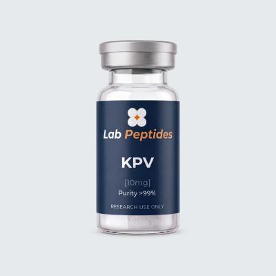
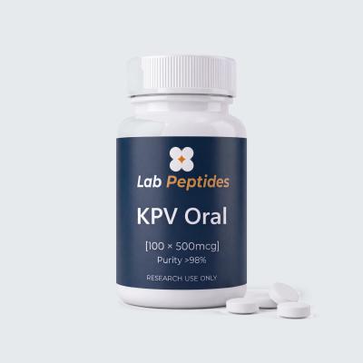
No published study directly compares the bioavailability of oral and subcutaneous KPV. The choice therefore depends on the tissue under study: the gut for oral protocols, systemic inflammation or tissues outside the gut for injectable ones. Lyophilised KPV 10 mg covers injectable protocols and solution preparation; oral KPV 500 mcg capsules suit digestive protocols. For the wider question of oral peptide absorption, see our article on oral peptides and bioavailability.
KPV vs BPC-157: two different gut profiles
KPV and BPC-157 are often mentioned together for digestive research. Their profiles are very different.
| Criterion | KPV | BPC-157 |
|---|---|---|
| Size | 3 amino acids | 15 amino acids (pentadecapeptide) |
| Origin | C-terminal fragment of α-MSH | Sequence derived from a protein in human gastric juice |
| Main mechanism described | NF-κB inhibition, uptake through PepT1 | Angiogenesis (VEGFR2), Akt-eNOS and nitric oxide pathway, ERK1/2 |
| Main research area | Colitis, skin and airway inflammation | Tendon, muscle and mucosal repair |
| Human data | No published trial | Three pilot studies according to McGuire et al., 2025 |
In short, KPV is studied to dampen inflammation, BPC-157 to support tissue repair. No published study has tested the two together. BPC-157 mechanisms are covered in our article BPC-157: molecular mechanisms and state of the science.
KPV, α-MSH and Melanotan: two ends of the same hormone
Melanotan 2 (MT-II) is a cyclic analogue of the 4-10 portion of α-MSH, built around the His-Phe-Arg-Trp motif that activates the MC1R, MC3R, MC4R and MC5R receptors. It tans the skin and acts on appetite and sexual function. KPV comes from the other end of the same hormone: positions 11 to 13, without the receptor-binding motif.
| Criterion | α-MSH | Melanotan 2 | KPV |
|---|---|---|---|
| Size | 13 amino acids | 7 residues, cyclic | 3 amino acids |
| His-Phe-Arg-Trp motif | Yes | Yes (D-Phe) | No |
| Pigmentation | Yes | Yes, strong | No |
| Anti-inflammatory | Yes | Yes, sensitive to the MC3/MC4 antagonist (Getting 2003) | Yes, insensitive to that antagonist (Getting 2003) |
KPV therefore lets researchers study the anti-inflammatory activity of α-MSH without its melanocortin effects. For receptor pharmacology, see our complete Melanotan 2 guide.
KPV dosage: concentrations and doses studied in the literature
No human dose has been established, since no clinical trial has been published. The amounts used in studies are:
- In vitro: 10 nM in most intestinal cell experiments (Dalmasso 2008); 0.1 to 10 µM on corneal cells (Bonfiglio 2006); 50 µg/mL on keratinocytes (Sung 2025).
- Oral, in mice: 100 µM in drinking water (Dalmasso 2008, Viennois 2016).
- Eye drops, in rabbits: 1, 5 or 10 mg/mL, 30 µL, two drops four times a day for 4 days (Bonfiglio 2006).
These values do not transfer directly from one species to another.
Reconstitution (research use only)
KPV 10 mg comes as a lyophilised powder. It is reconstituted with bacteriostatic water (sterile water with 0.9% benzyl alcohol). The volume of water sets the concentration, and therefore the amount held in each mark of a U-100 insulin syringe (100 units = 1 mL, 1 unit = 0.01 mL).
| Water added to the 10 mg vial | Concentration | 1 U-100 unit (0.01 mL) | 10 units (0.1 mL) |
|---|---|---|---|
| 1 mL | 10 mg/mL | 100 µg | 1 mg |
| 2 mL | 5 mg/mL | 50 µg | 500 µg |
| 4 mL | 2.5 mg/mL | 25 µg | 250 µg |
Example: with 2 mL of water, drawing 250 µg means 0.05 mL, i.e. 5 units on a U-100 syringe. Good practice: disinfect the stopper, run the water slowly down the vial wall, swirl gently without shaking, and wait for a clear solution.
Stability and storage
Lyophilised KPV is stored in the freezer (-20 °C), away from light and moisture, or in the fridge for near-term use. Once reconstituted, it is kept at 2-8 °C and used within the following weeks, with aseptic handling. No published study gives a precise stability window for KPV reconstituted in bacteriostatic water.
The main degradation pathway identified is cyclisation into Lys-Pro diketopiperazine, seen in acidic, alkaline or oxidising conditions (Pawar et al., 2015). Extreme pH and oxidising solutions should therefore be avoided. The same paper describes a validated HPLC method that separates KPV from its degradation products.
Safety and reported signals
Safety data are limited by the lack of human studies. In published animal and cell work:
- KPV nanoparticles did not affect intestinal cell viability or barrier function (Laroui 2010), and hyaluronic acid nanoparticles were described as non-toxic and biocompatible (Xiao 2017).
- KPV has no pigmenting effect, which sets it apart from α-MSH and Melanotan (Brzoska 2008, 2010).
- KPV did not reduce the ability of human neutrophils to kill the bacteria and yeast tested (Cutuli 2000), a classic concern with anti-inflammatory agents.
The absence of signals in animal studies is not proof of safety in humans.
Regulatory status
KPV is not an approved medicine in France, the European Union or the United States. In the US it was nominated for pharmacy compounding, placed in FDA Category 2 in September 2023 (substances raising significant safety risks), and the nomination was then withdrawn: the FDA page now lists it among substances "nominated but withdrawn", alongside BPC-157, Melanotan II and TB-500, stating that the agency has no human exposure data for KPV (page current as of 22 April 2026). In Europe, KPV is sold as a research reagent, not for human or veterinary use.
KPV peptide FAQ
What is the KPV peptide?
KPV is the tripeptide Lys-Pro-Val, fragment 11-13 of the hormone α-MSH. It keeps the hormone's anti-inflammatory activity without its pigmenting effect, and acts mainly by blocking the NF-κB pathway.
Are there human clinical trials of KPV?
No. No clinical trial has been published, and no study is registered under its name on ClinicalTrials.gov. The data come from cultured human cells and from mouse, rat and rabbit models.
Does oral KPV work?
In mice, yes for the gut: KPV dissolved in drinking water at 100 µM reduced DSS and TNBS colitis (Dalmasso 2008), via the PepT1 transporter. There are no human data on its oral absorption.
What KPV dosage has been studied?
No human dose has been established. Studies use 10 nM in vitro, 100 µM in drinking water in mice and 1 to 10 mg/mL eye drops in rabbits.
Does KPV tan the skin like Melanotan?
No. KPV lacks the His-Phe-Arg-Trp motif that activates MC1R on melanocytes. The absence of pigmentation is exactly why it was studied as an anti-inflammatory.
Conclusion
Three amino acids are enough to reproduce most of the anti-inflammatory activity of a thirteen-residue hormone, without its pigmenting effect. Its uptake through PepT1, which is overexpressed in the inflamed colon, makes KPV a tool of choice for colitis research. The data remain preclinical: no human trial has been published. It is a research reagent and should be studied as one.
Order your KPV 10 mg: lyophilised vial, stock in France, shipped from France. Complete your protocol with bacteriostatic water 10 mL.